

Lots of graphics on this page.... please be patient.
PRESS YOUR BROSER'S "BACK"
BUTTON TO RETURN TO THE PREVIOUS PAGE
| Sponges belong to the phylum Porifera, meaning pore bearing.
They really quite simple organisms, but vary greatly in both size and form.
Sponges are nearly exclusively marine. Some are solitary individuals,
and others occur in groups or colonies of individuals. They lack
internal organs and circulatory and digestive systems. External holes
or pores in the walls provide openings through which water carries food
and oxygen into the cells and a larger opening at the top, the osculum,
that permits the water to flow out. Sponges are primitive, simple,
multicellular, aquatic animals which first appeared in the Cambrian period,
some 570 million years ago, and have survived to the present day.
Fossil sponges are most common in the Cretaceous period. Sponges
vary in size from 1 cm (0.4 inches) to more than 1 meter (3.3 feet).
They vary greatly in shape, being commonly vase-shapes, such as Ventriculites;
spherical, such as Porosphaera; pear-shaped, such as Siphonia; leaf-shaped,
such as Elasmostoma; and branching, such as Doryderma.
Sponges may be either rigid solitary individuals or colonial, consisting
of many
|
|
 |
|
| The classification of sponges is primarily based on the
composition of the skeleton. In general, there are two kinds of cells
in the sponge body. One lines the internal cavity and which, by making
motions with hair-like flagella, creates food-carrying water currents.
The another forms the outer part of the body wall and sometimes secretes
skeletal structures of silica (siliceous), or calcium carbonate (calcareous),
called spicules. Some have skeletons comprised of many needle-like
single or multi-rayed spicules. These spicules may be fused together
to form a solid framework. The spicules are what give the sponge
it ability to be preserved as a fossil. Spicules from the fossils
of Cambrian sponges are indistinguishable from those of similar living
sponges.
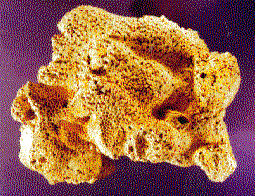
The natural bath sponge is not commonly preserved as a fossil because it lacks hard spicules, but Heliospongia had a preservable skeleton. Heliospongia had lumpy siliceous spicules that interlocked to form its thick cylindrical wall. The pores were present along the outside of the cylinder, and the osculum at the upper end. The Heliospongia is a sponge that does not bear much resemblance to what most people them of when the word sponge is mentioned. |
|
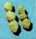
| This sponge has two pecularily-shaped fossils. These distinctive rows of bead-like spherical chambers are the calcareous skeletal remains of the sponge Girtyoceolia. The entire animal, of which only small parts are seen here, lived in shallow clear water, attached to the bottom. They branched upward from the base, and bore numerous spout-like openings on the outer walls of the chambers through which water currents passed, bringing in food and oxygen and carrying away wastes |  |
 |
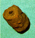
This is the sponge Amblysiphonella. It is similar in structure to Girtyocoelia, but differs in that the chambers are less bead-like or globular, and it has an axial tube seen as a circular aperture in the specimen. This specimen is rust-colored because the original skeletal material has been replaced by an iron-rich mineral. Amblysiphonella is restricted to Upper Pennsylvanian rocks, such as Topeka Limestone |
| The fossil record of sponges is not abundant, except in
a few scattered localities. Some
fossil sponges have worldwide distribution, while others are restricted to certain areas. Sponge fossils such as Hydnoceras and Prismodictya are found in the Devonian rocks of New York state. In Europe, the Jurassic limestone of the Swabian Alps are composed largely of sponge remains, some of which are well preserved. Many sponges are found in the Cretaceous Lower Greensand and Chalk formations of England, and in rocks from the upper part of the Cretaceous period in France. A famous locality for fossil sponges is the Cretaceous Farringdon Sponge Gravels in Farringdon, Oxfordshire in England. To see our collection of fossil sponges, just click on the icon below. |
|
|
|
|